
The Human Anatomy and Physiology Lab Manual is a comprehensive guide designed for undergraduate students, covering foundational topics like histology, skeletal systems, and physiological processes. It includes detailed visual aids, practical experiments, and step-by-step instructions to enhance hands-on learning and reinforce theoretical concepts. The manual emphasizes applied learning through interactive exercises and real-world applications, making it an essential resource for understanding human body structures and functions effectively.
1.1 Overview of the Lab Manual Structure
The lab manual is organized into clear, logical sections, each focusing on specific anatomical and physiological systems. It begins with foundational concepts, progressing to detailed explorations of histology, skeletal, muscular, nervous, and circulatory systems. Each chapter includes objectives, materials lists, step-by-step procedures, and review questions to reinforce learning. Visual aids like diagrams and micrographs are integrated throughout to enhance understanding. The structure is designed to align with course curricula, ensuring a systematic and comprehensive approach to hands-on learning.
Practical experiments and activities are balanced with theoretical explanations, encouraging students to apply knowledge in real-world contexts. The manual’s clear layout and concise instructions make it user-friendly, allowing learners to focus on mastering essential skills and concepts effectively.
1.2 Importance of Anatomy and Physiology in Lab Settings
Understanding anatomy and physiology is crucial in lab settings as it provides a foundation for analyzing bodily structures and their functions. This knowledge enables precise identification of tissues, cells, and systems during experiments. It aids in conducting accurate dissections, microscopic examinations, and physiological measurements. Additionally, it enhances critical thinking and problem-solving skills, essential for interpreting experimental results. In healthcare and research, anatomy and physiology form the basis for diagnostics, treatments, and understanding disease mechanisms, making them indispensable in both educational and professional lab environments.
1.3 Essential Tools and Equipment for Lab Experiments
The lab manual emphasizes the use of microscopes for examining tissue samples and cellular structures. Dissection tools, such as scalpels and forceps, are vital for exploring anatomical specimens. Slide preparation equipment, including microtomes and stains, aids in creating samples for histological analysis. Safety gear like gloves and goggles ensures protection during experiments; Additionally, measurement devices, such as blood pressure monitors and pH meters, are used to study physiological processes. These tools are essential for conducting accurate and safe lab experiments, fostering hands-on learning and practical understanding of anatomy and physiology.
Histology and Microscopic Anatomy
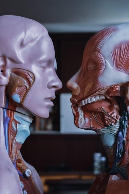
Histology and microscopic anatomy focus on studying tissues and cells under a microscope. This section explores tissue preparation, cellular structure identification, and understanding the microscopic organization of body tissues.
2.1 Preparing and Examining Tissue Samples
Preparing and examining tissue samples is a fundamental skill in histology. This involves fixing tissues to preserve structure, sectioning them into thin slices using microtomes, and staining to enhance visibility under a microscope. Proper techniques ensure clear cellular details, aiding in accurate tissue identification. Virtual labs and digital microscopy tools further support interactive learning, enabling students to analyze tissue samples remotely. This process is essential for understanding the microscopic organization of epithelial, connective, and muscle tissues, forming the basis of histological studies.
2.2 Understanding Epithelial, Connective, and Muscle Tissues
Epithelial tissues form protective layers, such as skin and lining of organs, while connective tissues provide support and connect structures, like bones and cartilage. Muscle tissues enable movement through contraction. Histological studies involve identifying these tissues under a microscope, noting characteristics like cell arrangement and fiber types. Lab exercises, including staining and imaging, help differentiate tissue types and understand their functional roles in maintaining body integrity and facilitating movement. This knowledge is vital for analyzing tissue samples and diagnosing conditions related to tissue structure and function.
2.3 Identifying Cellular Structures Under a Microscope
Identifying cellular structures under a microscope involves understanding tissue composition and using staining techniques to enhance visibility. Students learn to distinguish epithelial, connective, and muscle tissues, focusing on unique features like cell shape, arrangement, and fiber density. Practical exercises include preparing slides, adjusting microscope settings, and recognizing key structures such as nuclei, mitochondria, and cell membranes. This skill is essential for analyzing tissue samples, understanding cellular function, and diagnosing abnormalities. Microscopic observation is a cornerstone of anatomy and physiology studies, enabling detailed insights into the human body’s intricate structures.

Skeletal and Muscular Systems
This section provides a comprehensive exploration of the skeletal and muscular systems, focusing on bones, joints, and muscle structure. Interactive activities enhance understanding of movement, support, and function.
3.1 Identifying Bones and Joints in the Human Skeleton
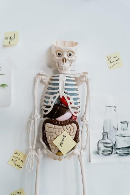
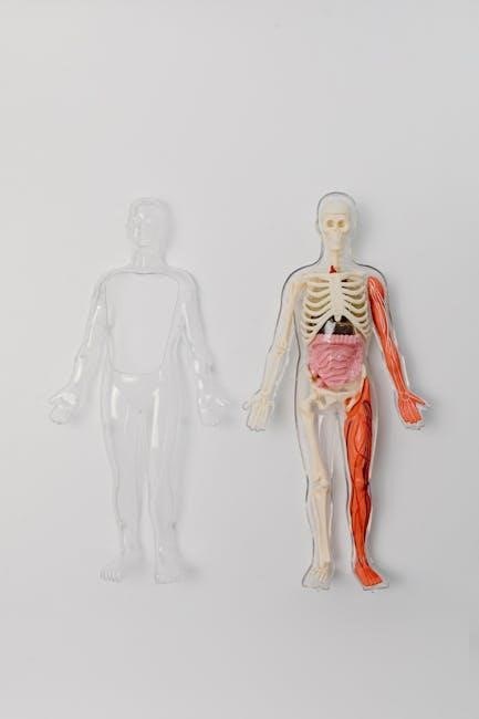
The human skeleton consists of 206 bones, categorized into long, short, flat, irregular, and sesamoid bones. This section guides learners in identifying and classifying bones based on their shapes and functions. It also explores the different types of joints, such as synovial, cartilaginous, and fibrous, and their roles in facilitating movement. Through detailed diagrams and hands-on activities, students learn to recognize anatomical landmarks and understand how joints contribute to mobility and structural support in the body. Practical exercises include labeling bones and analyzing joint structures to enhance comprehension of skeletal anatomy.
3.2 Examining Muscle Structure and Function
Muscles are dynamic tissues responsible for movement, support, and maintaining posture. This section focuses on the three types of muscle tissue: skeletal, smooth, and cardiac. Skeletal muscles are voluntary and attached to bones, enabling movement. Smooth muscles are involuntary, found in internal organs, and facilitate processes like digestion. Cardiac muscle is specialized for the heart, ensuring continuous, rhythmic contractions. Lab activities include microscopic examination of tissue samples and physiological experiments to understand muscle contraction mechanisms, emphasizing the role of motor neurons and muscle fibers in movement and overall bodily functions.
3.3 Lab Activities for Understanding Movement and Support
Lab activities focus on exploring the musculoskeletal system’s role in movement and support. Students analyze joint structures, observe muscle contractions, and study bone density. Practical exercises include dissecting skeletal muscles, measuring muscle strength, and simulating joint movements to understand biomechanics. These hands-on experiments enhance comprehension of how bones, muscles, and connective tissues collaborate to enable movement and provide structural support. Interactive models and real-world applications further reinforce the principles of human anatomy and physiology in maintaining posture and facilitating motion. This section emphasizes experiential learning to bridge theory with practice effectively.

Nervous and Digestive Systems
This section explores the intricate functions of the nervous and digestive systems through hands-on lab activities. Students examine neuron structures, analyze reflex mechanisms, and investigate digestion processes. Practical experiments include studying brain anatomy, observing muscle contractions in the digestive tract, and conducting simulations of nerve impulse transmission. These exercises provide a comprehensive understanding of how these systems maintain bodily functions and overall health.
4.1 Exploring the Structure of Neurons and Reflexes
This section delves into the anatomy of neurons, emphasizing their role as the building blocks of the nervous system. Students learn to identify dendrites, cell bodies, and axons under a microscope. Practical exercises involve tracing nerve pathways and simulating reflex actions. Hands-on activities include dissecting nerve tissue and observing synaptic transmission. The lab manual also explores reflex arcs, from sensory receptors to motor responses, highlighting the neural mechanisms behind involuntary actions. These exercises provide a foundational understanding of how neurons communicate and regulate bodily functions.
4.2 Analyzing the Brain and Spinal Cord
This section focuses on the detailed examination of the brain and spinal cord, highlighting their structural and functional roles. Students explore brain regions such as the cerebral cortex, cerebellum, and brainstem, analyzing their responsibilities in motor control, cognition, and sensory processing. The spinal cord’s segments and their connection to peripheral nerves are also studied. Practical activities include identifying anatomical features like meninges and ventricles, as well as understanding the spinal cord’s role in reflexes. These exercises provide a deeper understanding of the central nervous system’s complexity and its essential functions in controlling the body.
4.3 Investigating the Digestive Process and Nutrient Absorption
This section delves into the mechanisms of digestion and nutrient absorption, focusing on the roles of key organs like the stomach, small intestine, pancreas, and liver. Students explore how mechanical and chemical digestion break down food into absorbable nutrients. Practical exercises include simulating enzymatic reactions and observing nutrient absorption processes under a microscope. The section emphasizes understanding the physiology of digestion, including the role of hormones and the absorption of vitamins and minerals, to appreciate how the body sustains energy and growth through this complex system.
Cardiovascular and Respiratory Systems
This section explores the interdependent functions of the heart, blood vessels, and lungs, focusing on blood circulation, gas exchange, and oxygen delivery. Practical experiments and dissections reveal how these systems maintain life-sustaining processes, emphasizing the importance of physiological balance and homeostasis in the human body.
5.1 Studying Heart Anatomy and Blood Circulation
This section delves into the detailed anatomy of the heart, including its chambers, valves, and blood vessels. Students explore the pathways of blood circulation, from arterial to venous systems, and analyze the roles of the atria and ventricles in pumping blood. Through dissections, simulations, and microscopic observations, learners gain a deeper understanding of how the heart maintains blood flow and regulates pressure. Practical exercises emphasize the importance of cardiovascular health and its impact on overall bodily functions.
5.2 Examining the Lungs and Breathing Mechanisms
This section focuses on the structure and function of the lungs, including the bronchi, alveoli, and pleural membranes. Students explore the mechanics of breathing, such as inhalation and exhalation, and the role of the diaphragm in respiratory processes. Lab activities involve dissecting lung specimens, observing alveolar tissue under microscopes, and conducting simulations of gas exchange. The importance of pH balance in respiratory fluids and its impact on oxygen-carbon dioxide exchange is also emphasized, providing a comprehensive understanding of respiratory physiology and its vital role in maintaining homeostasis.
5.3 Conducting Experiments on Blood Pressure and Gas Exchange
Students perform hands-on experiments to measure blood pressure using sphygmomanometers and analyze factors affecting it, such as resistance and vessel diameter. Activities also involve simulating gas exchange in the lungs, observing oxygen and carbon dioxide diffusion across alveolar membranes. Practical exercises include monitoring pH levels in respiratory fluids and their impact on gas exchange efficiency. These experiments provide insights into cardiovascular and respiratory interactions, emphasizing the importance of maintaining homeostasis for proper bodily functions and overall health.